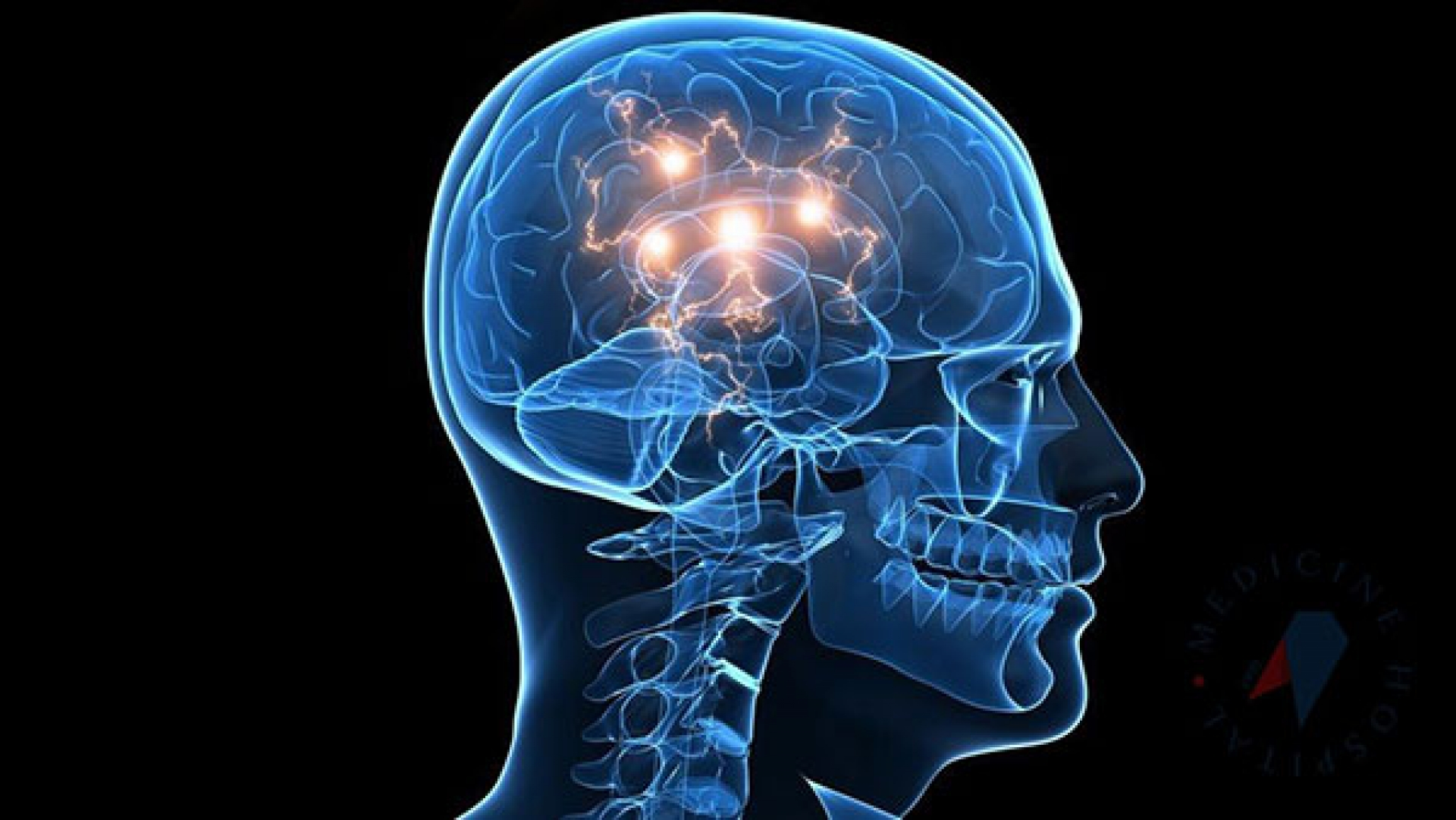


Arachnoid cysts are fluid-filled sacs that form within or between the thin membranes called arachnoid mater, which cover the brain and spinal cord. These cysts typically contain cerebrospinal fluid (CSF) and are most often congenital. In most cases, they do not cause symptoms, but large cysts may press on surrounding tissues and lead to clinical manifestations.
Etiology
Congenital: Caused by a developmental defect of the arachnoid membrane during the embryonic period (most common).
Acquired: May develop later due to trauma, infection (e.g., meningitis), surgery, or other causes.
According to Location
Cranial Arachnoid Cysts:
Most commonly found in the temporal lobe or the middle cranial fossa.
Spinal Arachnoid Cysts:
Can occur anywhere along the spinal cord.
Symptoms
Small arachnoid cysts often do not cause any symptoms and are discovered incidentally. However, large cysts or those compressing adjacent structures may lead to the following symptoms:
Cranial Cysts:
Headache.
Seizures (epilepsy).
Visual or hearing problems.
Balance disorders.
Hydrocephalus (increased intracranial pressure).
Spinal Cysts:
Back or neck pain.
Numbness and weakness in the arms or legs.
Bladder and bowel control problems.
Diagnosis
Magnetic Resonance Imaging (MRI): Used to evaluate the location, size, and effects of the arachnoid cyst on surrounding structures.
Computed Tomography (CT): Can assess the cyst’s content and any changes in the surrounding bone.
Treatment
Observation:
Regular monitoring may be sufficient for asymptomatic and non-growing cysts.
Surgical Intervention (For symptomatic or large cysts):
Cyst Fenestration: Draining the cyst and creating a connection with the surrounding CSF circulation.
Shunt Placement: Redirecting the cyst fluid to another body cavity (e.g., the abdominal cavity).
Prognosis
Asymptomatic cysts usually do not cause problems throughout life.
Symptomatic cysts that are treated often have good outcomes, but neurological recovery depends on the extent of damage caused to surrounding tissues.