Arteriovenous Malformations (AVMs)
Arteriovenous malformations (AVMs) are congenital vascular anomalies characterized by abnormal, direct connections between arteries and veins without the presence of a normal capillary network. This structure leads to both weakened vessel walls and abnormal blood flow.
Characteristics
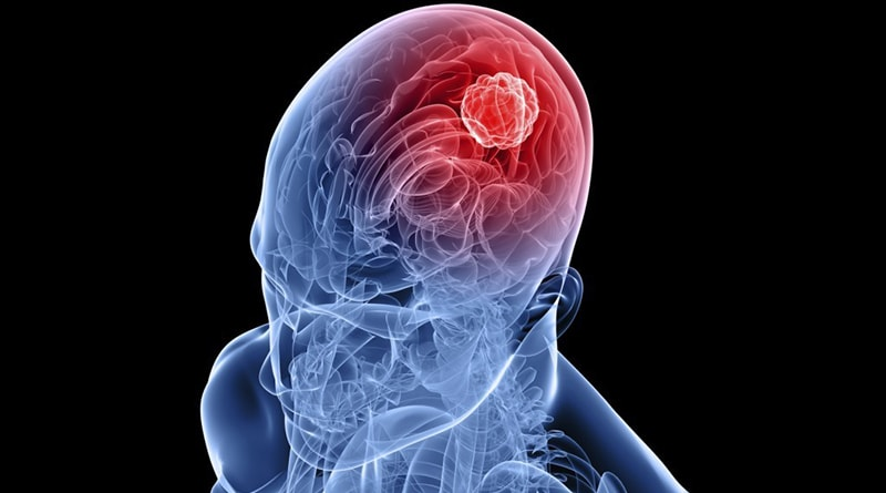
Most commonly found in the brain and spinal cord.
May grow over time or remain stable.
Carry a risk of bleeding; cerebral AVMs, in particular, can cause sudden stroke or even death.
Symptoms
Bleeding (often the first symptom)
Seizures
Headaches
Neurological deficits (weakness, speech impairment, sensory loss)
Some patients may be asymptomatic (discovered incidentally).
Diagnosis
Magnetic Resonance Imaging (MRI) and Computed Tomography (CT): Reveal structural abnormalities and possible bleeding.
Digital Subtraction Angiography (DSA): Considered the gold standard; provides detailed visualization of AVM vascular anatomy and flow characteristics.
Treatment
Surgical resection: Suitable AVMs are directly removed.
Endovascular embolization: A catheter is used to access and block AVM vessels.
Stereotactic radiosurgery (e.g., Gamma Knife): Radiation is used to close off small AVMs.
The choice of treatment depends on the AVM’s size, location, and risk of bleeding.
Conclusion
Untreated AVMs may lead to bleeding and neurological deterioration over time. Early intervention significantly reduces the risk of complications.
Would you like this formatted as a medical report or for a presentation?