Hydrocephalus is a condition that occurs as a result of an excessive accumulation of cerebrospinal fluid (CSF) around the brain and spinal cord. The excessive buildup of this fluid increases intracranial pressure, which can damage brain tissue.
Normal Function of Cerebrospinal Fluid (CSF)
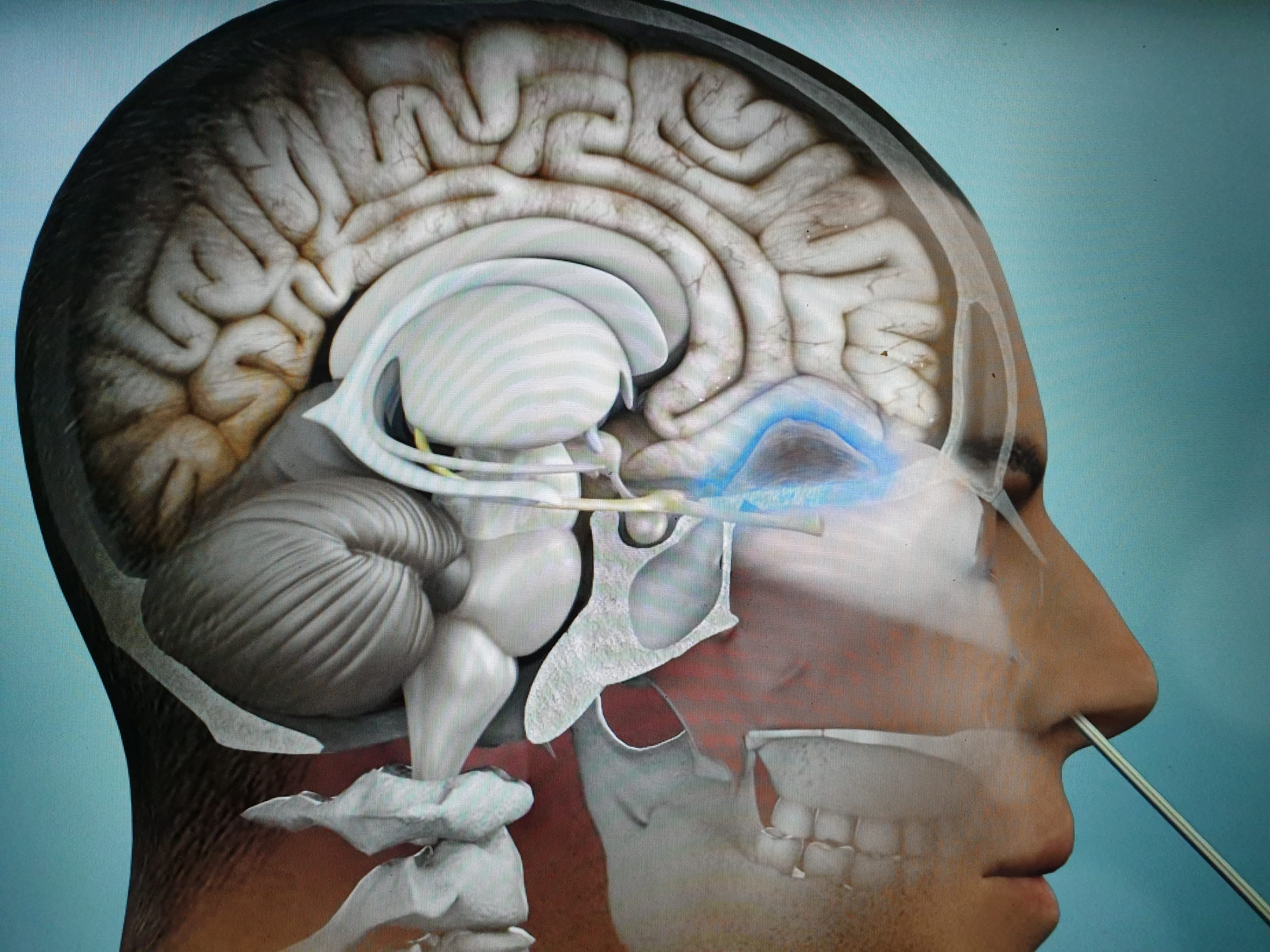
Production: CSF is produced by the choroid plexus in the brain ventricles.
Circulation: It circulates between the ventricles and the meninges.
Absorption: It is reabsorbed into the vascular system.
Hydrocephalus occurs due to problems in the production, circulation, or absorption of CSF in this system.
Classification
Congenital Hydrocephalus: Hydrocephalus present at birth. It is related to genetic anomalies or fetal developmental issues.
Acquired Hydrocephalus: Develops after birth.
Trauma
Infections: Such as meningitis.
Tumors: Can block CSF circulation.
According to Pathophysiology:
Obstructive (Non-communicating): There is a blockage in the flow of CSF between the ventricles or outward.
Communicating: CSF can circulate, but there is a problem with the absorption mechanism.
Normal Pressure Hydrocephalus (NPH): Typically seen in the elderly, CSF accumulation leads to a significant increase in intracranial pressure.
Symptoms
In Infants:
Abnormal head enlargement (bulging fontanel).
Downward gaze (sunsetting sign).
Irritability, difficulty feeding.
In Adults:
Headache, nausea, vomiting.
Blurred vision.
Gait disturbance, balance issues.
Memory and cognitive dysfunction (in NPH, the "gait difficulty, dementia, urinary incontinence" triad).
Diagnostic Methods
Neurological Examination
Imaging:
Magnetic Resonance Imaging (MRI): Used to assess the brain ventricles and fluid flow.
Computed Tomography (CT): Shows ventricular enlargement.
CSF Pressure Measurement: Done via lumbar puncture, particularly useful in diagnosing NPH.
Treatment
Surgical Interventions:
Ventriculoperitoneal (VP) Shunt: Directs CSF into the abdominal cavity.
Ventriculostomy: An alternative fluid pathway is created between the ventricles using an endoscopic method (ETV).
Medical Treatment: Provides temporary relief, usually used as preparation for surgery.
Complications
If left untreated, there is a risk of brain tissue damage, mental retardation, physical disability, or death.
Shunt Complications: Blockage, infection, overdrainage.
Prognosis
With early diagnosis and treatment, many patients can lead a normal life. However, regular follow-up is necessary due to the risks of complications.